Scratched into the Past

Greying apparitions
scurry silently
down hirsute paths,
nestled amongst
the cloying warmth
of flaking skin.
Their crude cement
seeps
across the contours
of our ancient scalps,
fixing oval shells
between seams
of folded flesh
and swaying stalks.

In search of lives
once lived
we comb through
fraying manes,
our past preserved
in bonds more fierce
than bone
or tooth
or claw.
Tenderly we run
fingers through hair,
tracing histories
to the withered stem
of every root.

A mummified adult man of the Ansilta culture, from the Andes of San Juan, Argentina, dating back approximately 2,000 years (Image Credit: Universidad Nacional de San Juan).

This poem is inspired by recent research, which has found that head lice can help us to analyse the remains of our ancestors.

When examining the DNA of our ancestors, scientists have tended to extract samples from the dense bone of the skull or from inside teeth. However, these are not always available, and it can be unethical or against cultural beliefs to take these samples from indigenous early remains. Such destructive sampling methods also cause severe damage to the specimens that can compromise future scientific analysis. As such, it is necessary to develop alternative techniques that enable us to analyse the remains of our ancestors in a less invasive manner. In this new study, researchers have demonstrated how head lice and their eggs can be used to provide a potential solution to this problem.

Head lice are tiny insects that live in hair, and nits are the empty egg cases attached to hair that head lice hatch from. Most ancient humans carried head lice, and their nits can often be found in historical hair specimens. In this new study, researchers found that human DNA can be extracted from the ‘cement’ that head lice used to glue nits to human hairs. By examining this nit-cement from a number of mummified remains the researchers were able to show that it contained the same concentration of DNA as a tooth, and twice as much as from a bone. Some of the mummies that were examined in this study came from Calingasta, a region from the province of San Juan in Argentina, and by comparing their nit-cement-extracted DNA to samples from other specimens, the researchers were able to show that the original peoples of Calingasta migrated from Amazonia about 2,000 years ago, a journey of over 5,000 km. The results from this study thereby demonstrate that this nit-cement can be used to enable more samples to be studied from human remains, especially in cases where bone and tooth samples are unavailable.


from ScienceBlog.com https://ift.tt/3eJxZcE

Leveraging space to advance stem cell science and medicine

The secret to producing large batches of stem cells more efficiently may lie in the near-zero gravity conditions of space. Scientists at Cedars-Sinai have found that microgravity has the potential to contribute to life-saving advances on Earth by facilitating the rapid mass production of stem cells.

new paper, led by Cedars Sinai and published in the peer-review journal Stem Cell Reports, highlights key opportunities discussed during the 2020 Biomanufacturing in Space Symposium to expand the manufacture of stem cells in space.

Biomanufacturing—a type of stem cell production that uses biological materials such as microbes to produce substances and biomaterials suitable for use in preclinical, clinical, and therapeutic applications—can be more productive in microgravity conditions.

“We are finding that spaceflight and microgravity is a desirable place for biomanufacturing because it confers a number of very special properties to biological tissues and biological processes that can help mass produce cells or other products in a way that you wouldn’t be able to do on Earth,” said stem cell biologist Arun Sharma, PhD, research scientist and head of a new research laboratory in the Cedars-Sinai Board of Governors Regenerative Medicine Institute, Smidt Heart Institute and Department of Biomedical Sciences.

“The last two decades have seen remarkable advances in regenerative medicine and exponential advancement in space technologies enabling new opportunities to access and commercialize space,” he said.

Attendees at the virtual space symposium in December identified more than 50 potential commercial opportunities for conducting biomanufacturing work in space, according to the Cedars-Sinai paper. The most promising fell into three categories: disease modeling, biofabrication, and stem-cell-derived products.

The first, disease modeling, is used by scientists to study diseases and possible treatments by replicating full-function structures—whether using stem cells, organoids (miniature 3D structures grown from human stem cells that resemble human tissue), or other tissues.

Investigators have found that once the body is exposed to low-gravity conditions for extended periods of time, it experiences accelerated bone loss and aging. By developing disease models based on this accelerated aging process, research scientists can better understand the mechanisms of the aging process and disease progression.

“Not only can this work help astronauts, but it can also lead to us manufacturing bone constructs or skeletal muscle constructs that could be applied to diseases like osteoporosis and other forms of accelerated bone aging and muscle wasting that people experience on Earth,” said Sharma, who is the corresponding author of the paper.

Another highly discussed topic at the symposium was biofabrication, which uses manufacturing processes to produce materials like tissues and organs. 3D printing is one of the core biofabrication technologies.

A major issue with producing these materials on Earth involves gravity-induced density, which makes it hard for cells to expand and grow. With the absence of gravity and density in space, scientists are hopeful that they can use 3D printing to print unique shapes and products, like organoids or cardiac tissues, in a way that can’t be replicated on Earth.

The third category has to do with the production of stem cells and understanding how some of their fundamental properties are influenced by microgravity. Some of these properties include potency, or the ability of a stem cell to renew itself, and differentiation, the ability for stem cells to turn into other cell types.

Understanding some of the effects of spaceflight on stem cells can potentially lead to better ways to manufacture large numbers of cells in the absence of gravity. Scientists from Cedars-Sinai will be sending stem cells into space early next year, in conjunction with NASA and a private contractor, Space Tango, to test whether it is possible to produce large batches in a low gravity environment.

“While we are still in the exploratory phase of some of this research, this is no longer in the realm of science fiction,” Sharma said. “Within the next five years we may see a scenario where we find cells or tissues that can be made in a way that is simply not possible here on Earth. And I think that’s extremely exciting.”


from ScienceBlog.com https://ift.tt/34bIqUB

How cells make curved mouth to ‘eat’

A new study shows how cell membranes curve to create the “mouths” that allow the cells to consume things that surround them.

“Just like our eating habits basically shape anything in our body, the way cells ‘eat’ matters for the health of the cells,” said Comert Kural, associate professor of physics at The Ohio State University and lead author of the study. “And scientists did not, until now, understand the mechanics of how that happened.”

The study, published recently in the journal Developmental Cell, found that the intercellular machinery of a cell assembles into a highly curved basket-like structure that eventually grows into a closed cage. Scientists had previously believed that structure began as a flat lattice.

Membrane curvature is important, Kural said: It controls the formation of the pockets that carry substances into and out of a cell.

The pockets capture substances around the cell, forming around the extracellular substances, before turning into vesicles – small sacs one-one millionth the size of a red blood cell. Vesicles carry important things for a cell’s health – proteins, for example – into the cell. But they can also be hijacked by pathogens that can infect cells.

But the question of how those pockets formed from membranes that were previously believed to be flat had stymied researchers for nearly 40 years.

“It was a controversy in cellular studies,” Kural said. “And we were able to use super-resolution fluorescence imaging to actually watch these pockets form within live cells, and so we could answer that question of how they are created.

“Simply put, in contrast to the previous studies, we made high-resolution movies of cells instead of taking snapshots,” Kural said. “Our experiments revealed that protein scaffolds start deforming the underlying membrane as soon as they are recruited to the sites of vesicle formation.”

That contrasts with previous hypotheses that the protein scaffolds of a cell had to go through an energy-intensive reorganization in order for the membrane to curve, Kural said.

The way cells consume and expel vesicles plays a key role for living organisms. The process helps clear bad cholesterol from blood; it also transmits neural signals. The process is known to break down in several diseases, including cancer and Alzheimer’s disease.

“Understanding the origin and dynamics of membrane-bound vesicles is important – they can be utilized for delivering drugs for medicinal purposes but, at the same time, hijacked by pathogens such as viruses to enter and infect cells,” Kural said. “Our results matter, not only for our understanding of the fundamentals of life, but also for developing better therapeutic strategies.”

Emanuele Cocucci, an assistant professor in Ohio State’s College of Pharmacy, co-authored this study, along with researchers from UC Berkeley, UC Riverside, Iowa State University, Purdue University and the Chinese Academy of Sciences.


from ScienceBlog.com https://ift.tt/3eC1flI

Biomarkers for PTSD identified by Russian researchers

War veterans have helped scientists of South Ural State University identify biological indicators which signal whether people have a post-traumatic stress disorder or a susceptibility to it. The research results are published in the Journal of Psychiatric Research, a top-rated scientific journal.

In their research works on studying post-traumatic stress disorder, the representatives of the SUSU School of Medical Biology have reached a new level. Earlier, they used an experimental method of stimulating post-traumatic stress disorder in rats and tracking changes in their bodies. Now, the world scientific community has been presented the results of the clinical study.

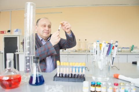
The work began back in 2016. More than a hundred people became participants: patients of the Chelyabinsk Regional Clinical Therapeutic Hospital for War Veterans and combat action participants who had not been diagnosed with the disorder. The scientists conducted clinical interviews with each participant and used diagnostic scales to assess the severity of the condition. At the same time, blood samples were taken to compare the changes in the body and determine the markers of PTSD.

“All participants of the research were separated from traumatic events by a long time interval: at least ten years. We managed to find out that even after a long time, the levels of cortisol and gamma-aminobutyric acid differed from the reference values. Gamma-aminobutyric acid appeared to be the most sensitive indicator, it can be considered as a marker of a long-lasting PTSD. A low acid level was demonstrated in a group of people susceptible to stress,” said Doctor of Sciences (Biology), Director of the SUSU School of Medical Biology Vadim Tseylikman.

All of those who had experienced traumatic events had elevated cortisol levels. It was this discovery that allowed the researchers to once again prove that despite extensive experimental studies of PTSD, the structure of the disease is not homogeneous.

“Thanks to this research, conducted by the Molecular Genetic Studies of Health and Well-Being Laboratory headed by Inna Feklicheva and by our colleagues from the Netherlands, Dr. Marco Boks and Dr. Ron de Kloet, we are now able to predict whether a person would be susceptible to PTSD or resistant to the disease after combat stress judging by the changes in blood plasma,” added Vadim Tseylikman.

The international team of scientists intends to continue the work and now consider changes in the genomic profile of people who have experienced traumatic events. The remaining blood samples will be examined. The SUSU’s consortium partner, the Institute of Cytology and Genetics of Siberian Branch of the Russian Academy of Sciences, will be engaged in this work.

Thanks to the research works by the School of Medical Biology, SUSU entered the RUR world ranking in the subject area of Medical Sciences and was listed in the top ten among Russian universities.

South Ural State University (SUSU) is a university of digital transformations, where innovative research is conducted in most of the priority fields of science and technology development. In accordance with the strategy of scientific and technological development of the Russian Federation, the university is focused on the development of big scientific interdisciplinary projects in the field of digital industry, materials science, and ecology. In the Year of Science and Technology, SUSU has become the winner in the competition under the Priority-2030 program. The university acts as a regional project office of the World-class Ural Interregional Research and Education Centre (UIREC).


from ScienceBlog.com https://ift.tt/3qE3Scn

COVID-19 can trigger self-attacking antibodies

Infection with the virus that causes COVID-19 can trigger an immune response that lasts well beyond the initial infection and recovery—even among people who had mild symptoms or no symptoms at all, according to Cedars-Sinai investigators. The findings are published in the Journal of Translational Medicine.

When people are infected with a virus or other pathogen, their bodies unleash proteins called antibodies that detect foreign substances and keep them from invading cells. In some cases, however, people produce autoantibodies that can attack the body’s own organs and tissues over time.

The Cedars-Sinai investigators found that people with prior infection with SARS-CoV-2, the virus that causes COVID-19, have a wide variety of autoantibodies up to six months after they have fully recovered. Prior to this study, researchers knew that severe cases of COVID-19 can stress the immune system so much that autoantibodies are produced. This study is the first to report not only the presence of elevated autoantibodies after mild or asymptomatic infection, but their persistence over time.

“These findings help to explain what makes COVID-19 an especially unique disease,” said Justyna Fert-Bober, PhD, research scientist in the Department of Cardiology at the Smidt Heart Institute and co-senior author of the study. “These patterns of immune dysregulation could be underlying the different types of persistent symptoms we see in people who go on to develop the condition now referred to as long COVID-19.”

To conduct their study, the Cedars-Sinai research team recruited 177 people with confirmed evidence of a previous infection with SARS-CoV-2. They compared blood samples from these individuals with samples taken from healthy people prior to the pandemic. All those with confirmed SARS-CoV-2 infection had elevated levels of autoantibodies. Some of the autoantibodies also have been found in people with diseases in which the immune system attacks its own healthy cells, such as lupus and rheumatoid arthritis.

“We found signals of autoantibody activity that are usually linked to chronic inflammation and injury involving specific organ systems and tissues such as the joints, skin and nervous system,” said Susan Cheng, MD, MPH, MMSc, director of the Institute for Research on Healthy Aging in the Department of Cardiology at the Smidt Heart Institute and co-senior author of the study.

Some of the autoantibodies have been linked to autoimmune diseases that typically affect women more often than men. In this study, however, men had a higher number of elevated autoantibodies than women.

“On the one hand, this finding is paradoxical given that autoimmune conditions are usually more common in females,” Fert-Bober said. “On the other hand, it is also somewhat expected given all that we know about males being more vulnerable to the most severe forms of COVID-19.”

The research team is interested in expanding the study to look for the types of autoantibodies that may be present and persist in people with long-haul COVID-19 symptoms. Because this study was in people infected before the advent of vaccines, the researchers will also examine whether autoantibodies are similarly generated in people with breakthrough infections.

“If we can better understand these autoantibody responses, and how it is that SARS-CoV-2 infection triggers and drives these variable responses, then we can get one step closer to identifying ways to treat and even prevent these effects from developing in people at risk,” Cheng said.

Journal of Translational Medicine


from ScienceBlog.com https://ift.tt/3eKMTPT

Speeding up directed evolution of molecules in the lab

Natural evolution is a slow process that relies on the gradual accumulation of genetic mutations. In recent years, scientists have found ways to speed up the process on a small scale, allowing them to rapidly create new proteins and other molecules in their lab.

This widely-used technique, known as directed evolution, has yielded new antibodies to treat cancer and other diseases, enzymes used in biofuel production, and imaging agents for magnetic resonance imaging (MRI).

Researchers at MIT have now developed a robotic platform that can perform 100 times as many directed-evolution experiments in parallel, giving many more populations the chance to come up with a solution, while monitoring their progress in real-time. In addition to helping researchers develop new molecules more rapidly, the technique could also be used to simulate natural evolution and answer fundamental questions about how it works.

“Traditionally, directed evolution has been much more of an art than a science, let alone an engineering discipline. And that remains true until you can systematically explore different permutations and observe the results,” says Kevin Esvelt, an assistant professor in MIT’s Media Lab and the senior author of the new study.

MIT graduate student Erika DeBenedictis and postdoc Emma Chory are the lead authors of the paper, which appears today in Nature Methods.

Rapid evolution

Directed evolution works by speeding up the accumulation and selection of novel mutations. For example, if scientists wanted to create an antibody that binds to a cancerous protein, they would start with a test tube of hundreds of millions of yeast cells or other microbes that have been engineered to express mammalian antibodies on their surfaces. These cells would be exposed to the cancer protein that the researchers want the antibody to bind to, and researchers would pick out those that bind the best.

Scientists would then introduce random mutations into the antibody sequence and screen these new proteins again. The process can be repeated many times until the best candidate emerges.

About 10 years ago, as a graduate student at Harvard University, Esvelt developed a way to speed up directed evolution. This approach harnesses bacteriophages (viruses that infect bacteria) to help proteins evolve faster toward a desired function. The gene that the researchers hope to optimize is linked to a gene needed for bacteriophage survival, and the viruses compete against each other to optimize the protein. The selection process is run continuously, shortening each mutation round to the lifespan of the bacteriophage, which is about 20 minutes, and can be repeated many times, with no human intervention needed.

Using this method, known as phage-assisted continuous evolution (PACE), directed evolution can be performed 1 billion times faster than traditional directed evolution experiments. However, evolution often fails to come up with a solution, requiring the researchers to guess which new set of conditions will do better.

The technique described in the new Nature Methods paper, which the researchers have named phage and robotics-assisted near-continuous evolution (PRANCE), can evolve 100 times as many populations in parallel, using different conditions.

In the new PRANCE system, bacteriophage populations (which can only infect a specific strain of bacteria) are grown in wells of a 96-well plate, instead of a single bioreactor. This allows for many more evolutionary trajectories to occur simultaneously. Each viral population is monitored by a robot as it goes through the evolution process. When the virus succeeds in generating the desired protein, it produces a fluorescent protein that the robot can detect.

“The robot can babysit this population of viruses by measuring this readout, which allows it to see whether the viruses are performing well, or whether they’re really struggling and something needs to be done to help them,” DeBenedictis says.

If the viruses are struggling to survive, meaning that the target protein is not evolving in the desired way, the robot can help save them from extinction by replacing the bacteria they’re infecting with a different strain that makes it easier for the viruses to replicate. This prevents the population from dying out, which is a cause of failure for many directed evolution experiments.

“We can tune these evolutions in real-time, in direct response to how well these evolutions are occurring,” Chory says. “We can tell when an experiment is succeeding and we can change the environment, which gives us many more shots on goal, which is great from both a bioengineering perspective and a basic science perspective.”

Novel molecules

In this study, the researchers used their new platform to engineer a molecule that allows viruses to encode their genes in a new way. The genetic code of all living organisms stipulates that three DNA base pairs specify one amino acid. However, the MIT team was able to evolve several viral transfer RNA (tRNA) molecules that read four DNA base pairs instead of three.

In another experiment, they evolved a molecule that allows viruses to incorporate a synthetic amino acid into the proteins they make. All viruses and living cells use the same 20 naturally occurring amino acids to build their proteins, but the MIT team was able to generate an enzyme that can incorporate an additional amino acid called Boc-lysine.

The researchers are now using PRANCE to try to make novel small-molecule drugs. Other possible applications for this kind of large-scale directed evolution include trying to evolve enzymes that degrade plastic more efficiently, or molecules that can edit the epigenome, similarly to how CRISPR can edit the genome, the researchers say.

With this system, scientists can also gain a better understanding of the step-by-step process that leads to a particular evolutionary outcome. Because they can study so many populations in parallel, they can tweak factors such as the mutation rate, size of original population, and environmental conditions, and then analyze how those variations affect the outcome. This type of large-scale, controlled experiment could allow them to potentially answer fundamental questions about how evolution naturally occurs.

“Our system allows us to actually perform these evolutions with substantially more understanding of what’s happening in the system,” Chory says. “We can learn about the history of the evolution, not just the end point.”

The research was funded by the MIT Media Lab, an Alfred P. Sloan Research Fellowship, the Open Philanthropy Project, the Reid Hoffman Foundation, the National Institute of Digestive and Kidney Diseases, the National Institute for Allergy and Infectious Diseases, and a Ruth L. Kirschstein NRSA Fellowship from the National Cancer Institute.


from ScienceBlog.com https://ift.tt/32uGYfH

Easy-to-take medicine better at suppressing HIV in children

A once-a-day antiretroviral medicine that is low-cost and easy for children to take is also more effective at suppressing HIV than standard treatments, according to a global trial led by researchers at UCL.

The study, published today in The New England Journal of Medicine, found that dolutegravir-based regimens, which are already widely used to treat adults, reduced the chances of treatment failure among young people aged three to 18 by around 40% compared to standard treatments.

The findings were based on a randomised controlled trial called ODYSSEY involving more than 700 children from 29 clinical centres in Africa, Europe and Asia, who were randomly given either dolutegravir or standard anti-HIV drugs, and who were followed up for at least two years.

The findings from the trial, which was sponsored by the Penta Foundation and funded by ViiV Healthcare, informed new guidance by the World Health Organisation, recommending the use of dolutegravir-based treatment for children.

Professor Diana Gibb (MRC Clinical Trials Unit at UCL), principal investigator of the ODYSSEY trial and one of the senior authors of the paper, said: “Our findings provide strong evidence for the global roll-out of dolutegravir for children with HIV.

“Medical treatments for children often lag woefully behind those of adults because of the separate formulations and studies that are needed. With the evidence from ODYSSEY which used simplified dosing, this treatment gap has been reduced and we hope that countries can quickly scale up children’s access to treatment globally.”

Lead author Dr Anna Turkova (MRC Clinical Trials Unit at UCL) said: “About 1.8 million children live with HIV but they have had limited treatment options, with medicines that taste unpalatable, that need to be taken twice a day, or that come in large pills that are difficult to swallow.

“Dolutegravir is given in small tablets usually once a day and the baby pills can be dispersed in water, meaning it’s a lot easier for young children to take. This is important in encouraging uptake of the treatment and adherence to it over many years. Sadly, only about half of children living with HIV are currently receiving treatment, and those who are not treated face high risks of impaired immunity and worsening health.”

In the study, researchers found that 14% of children receiving dolutegravir experienced treatment failure over two years compared to 22% of children receiving standard treatment. Treatment failure was deemed to occur if the virus became measurable in the blood – i.e. it was not fully suppressed – or if the child had symptoms of HIV-related ill health. Such a failure may be a result of the drug not being taken as well as the drug not working.

Evidence from adults shows dolutegravir has a high genetic barrier to resistance, meaning viruses are less likely to become resistant to it over time. This was replicated in the ODYSSEY trial, with much less resistance occurring among children and adolescents on dolutegravir-based treatment.

Past studies have suggested dolutegravir may be associated with weight gain among adults but the researchers said the new findings were reassuring for children, with those given dolutegravir gaining 1kg more and growing 1cm higher over two years – both indicating better growth rather than abnormal weight gain. Children in the dolutegravir arm had better lipid profiles, meaning lower risk of cardiovascular disease in the long term.

In the main trial, the children all weighed over 14kg and most were aged six and over. The therapy’s effectiveness was also looked at among young children and babies weighing under 14kg, enrolled as a separate group in the trial; results are yet to be published.

The trial participants were enrolled in Uganda, Zimbabwe, South Africa, Thailand, the UK, Spain, Portugal and Germany. Most of the participants were based in sub-Saharan Africa, where most children living with HIV are.

Earlier findings from the ODYSSEY trial showed that children weighing 20kg or more could safely take adult-strength tablets of dolutegravir, informing WHO dosing guidance and contributing to new licences for the drug in the United States and Europe during 2020.

Dr Cissy Kityo, from the Joint Clinical Research Centre in Uganda, the country enrolling most children into ODYSSEY, said: “Simplifying the dosing is crucial. Older children being able to take the same tablets as adults immediately opens access to dolutegravir for the majority of children living with HIV. It greatly simplifies procurement for national health systems in low and middle income countries and lowers costs.”

Dolutegravir is an integrase inhibitor – that is, it suppresses HIV by inhibiting integrase, an enzyme that the virus needs in order to replicate.


from ScienceBlog.com https://ift.tt/3eI7Ue1

With ‘Test-to-Stay,’ Children and Staff Can Safely Remain in School After COVID Exposures

Children and staff who repeatedly test negative for COVID-19 after contact with someone who has the illness can safely remain in school if universal masking programs are in place, according to a new “test-to-stay” study report from the ABC Science Collaborative.

The finding provides a safe alternative to quarantining people who have been exposed to COVID-19 and enables schools to remain open without interruptions. This research will be used by the N.C. Department of Health and Human Services to consider revising its guidelines on quarantine for schools across North Carolina.Du

The research, coming as the country faces the omicron variant, provides a more practical and focused approach to the “test-to-stay” protocols that the Centers for Disease Control and Prevention recently endorsed, which require testing of anyone within three feet of an infected person at school despite both parties being masked.

Watch Dr. Danny Benjamin discuss the findings on YouTube.

ABC Science researchers have raised concerns that this widespread “test-to-stay” approach is likely to overwhelm resource-limited schools and result in insurmountable logistical hurdles.

By contrast, the ABC Science Collaborative’s focused “test-to-stay” approach only requires testing if at least one of the exposed individuals is unmasked.

This focused approach reduces testing by ~80%; the transmission is still very low at 1.7%; and testing volume is such that most schools can complete the testing at each school, thereby serving more vulnerable children.

“The focused ‘test-to-stay’ protocol substantially reduced student absences from school after in-school exposure to COVID-19, keeping more kids in school and on a consistent educational routine,” said Danny Benjamin, MD, MPH, PhD, co-chair of the ABC Science Collaborative and Distinguished Professor of Pediatrics, Duke University. “Our research has taught us that ‘test-to-stay’ is a focused, practical way for children to avoid being out of the classroom after an exposure and can be a win-win strategy for keeping our children and schools safe without overwhelming the system.”

North Carolina schools and school districts were eligible to participate in the ABC Science study if they had a universal masking policy and approval from their local board of education and local health department. Individuals at participating schools were eligible if they were identified as a close contact by the local health department and were required to quarantine following an in-school, unmasked COVID-19 exposure.

These individuals could participate in “test-to-stay” if they were asymptomatic and consented to participate in the “test-to-stay” research study. Close contacts were given the option to quarantine according to local policies.

Participants in the study were given a SARS-CoV-2 rapid test at school when they were identified as a close contact and received testing every other day up to four times during the first seven days after the known exposure. Participants remained in school if they tested negative and were asymptomatic. A positive COVID-19 test or the development of symptoms on any day after exposure required isolation according to state public health guidelines.

Over the course of a six-week duration, more than 880 tests were performed among more than 360 participants.

“There were no instances in the ABC Science Collaborative ‘test-to-stay’ study where an exposed child became infected and went on to infect other children or adults within the school building,” said Kanecia Zimmerman, MD, MPH, co-chair of the ABC Science Collaborative and Test-to-Stay Principal Investigator, and a pediatrician at Duke University. “Implementation of ‘test-to-stay’ reduced missed days of school during quarantine by more than 90%, saving 1,628 days of in-person learning over the course of the study.”

This research was funded in part by the Rapid Acceleration of Diagnostics (RADx) Underserved Populations (RADx-UP); National Institutes of Health; the Trial Innovation Network, which is an innovative collaboration addressing critical roadblocks in clinical research and accelerating the translation of novel interventions into life-saving therapies; and the National Institute of Child Health and Human Development (NICHD).

For more information, visit https://abcsciencecollaborative.org.


from ScienceBlog.com https://ift.tt/3mKuLKr

Pregnant women living near fracking sites face higher risk of hypertension

In a study of nearly three million births over 13 years, Oregon State University researchers found that pregnant women living in close proximity to oil or gas drilling sites in Texas were more likely to have hypertension compared to those who lived farther away.

After accounting for other factors that influence high blood pressure, the results showed that pregnant women who lived within 1 kilometer of an active drilling site were 5% more likely to develop hypertension and 26% more likely to develop eclampsia, a more severe form of high blood pressure that can cause seizures and pose a serious risk to both mothers and infants.

These findings, published earlier this month in the International Journal of Epidemiology, align with a 2019 National Toxicology Program statement that air pollution is associated with an increased risk in hypertensive disorders.

“Oil and gas drilling produces air pollutions, so it made sense that we would find a similar increased risk near oil and gas sites,” said Mary Willis, lead author on the paper and a postdoctoral scholar in OSU’s College of Public Health and Human Sciences.

This was the first study to look specifically at the association between oil and gas drilling sites and hypertension during pregnancy. It examined birth certificates from more than 2.8 million babies born to mothers who lived less than 10 kilometers from an active or future drilling site in Texas during the years 1996-2009. Texas was chosen because it has the most oil and gas drilling activity of any state.

Birth certificates document risk factors experienced by the mother, including hypertension and eclampsia. The certificates included in the study did not record pre-eclampsia, a more common, milder form of eclampsia.

The increased odds of hypertension and eclampsia dissipated as mothers’ proximity lessened, with the effect dissipating entirely after 3 kilometers.

Nationwide, up to 8% of pregnancies are affected by hypertensive conditions and 16% of maternal deaths are attributed to complications arising from high blood pressure, the study authors note. Gestational hypertension may last for months after childbirth and costs the health care system over $1 billion annually.

In the U.S., an estimated 11.3 million people live within 1 km of a drilling site.

“In Texas, drilling sites can be as close as 45 meters from residences,” Willis said. “Most people think that no one lives near drilling, but there are large populations that live in close proximity to this industry. As the industry expands, this means that an increasing segment of the population may be vulnerable to drilling-related pollution.”

The study also accounted for the potential socioeconomic benefits of oil and gas drilling, which include more jobs and higher income for those living near a drilling site. These financial benefits may help mitigate some of the negative health effects of pollution associated with drilling sites, Willis said.

More research is needed to understand the precise relationship between oil and gas drilling sites and hypertensive disorders during pregnancy, Willis said. Drilling sites cause air pollution in the form of flaring and increased road traffic, along with water contamination and noise and light pollution. It’s unclear how those individual factors affect pregnancy, and at what point in the pregnancy mothers are most vulnerable.

Co-authors on the paper were Molly Kile and Perry Hystad in OSU’s College of Public Health and Human Sciences, Elaine Hill at the University of Rochester and Susan Carozza, recently retired from OSU.


from ScienceBlog.com https://ift.tt/32MRluM

‘Never seen anything as effective’ – the not-so-new-drug repurposed for a rare disease

The earliest signs of alkaptonuria are often subtle and harmless, like a diaper stained black. However, over the years, this rare genetic disease can lead to a lifetime of surgery. Now, after 20 years of research, a not-so-new drug can offer relief for thousands of patients worldwide.

The disease, also known as AKU, prevents the breakdown of a chemical called homogentisic acid in the body. The kidneys help to clear this chemical and get rid of it through urine. When exposed to the air, it turns black and this is how parents usually spot the first sign of the condition in children.

However, some of the homogentisic acid remains in the body and builds up slowly over time. This starts to cause damage in the areas that it accumulates, such as the cartilage and heart valves.

‘Similar symptoms appear in most patients, with spinal problems in their 20s or 30s, then severe joint deterioration during their 30s, 40s and 50s, and then heart problems in their 50s and later,’ said Nick Sireau, CEO of the AKU Society in the UK.

Sireau has two sons with AKU, which prompted his interest in the condition. AKU affects about one in a million people, who each have two defective copies of a gene called HGD. That means that Sireau’s sons inherited defective HGD genes from him and their mother, both of whom are genetic carriers with no symptoms.

‘We were quite fortunate because they were diagnosed at birth. Our eldest is 20 years old and our other son is 17,’ said Sireau. ‘The only symptoms they’ve really had is the urine going red-black.

‘For us, the parents, it has had obviously much more of an impact, because I’ve been now working on this for 17 years. It’s become my job.’

Tantalising

The AKU Society has helped to fund research into the condition, as there were no treatments available. Many patients need joint and heart valve replacement surgery as the symptoms progress. However, the promise of a new treatment has remained tantalisingly on the horizon for almost two decades, says Sireau.

‘When my first son was diagnosed, we went to Great Ormond Street (a children’s hospital in London, UK) and had a meeting with a consultant who said there was really not much we could do, but there was the potential of a treatment in the next 10 years or so but that it was very, very early stage. That’s when we heard about nitisinone and that the National Institutes of Health in America was starting to look at it for AKU.’

Nitisinone is a drug already in use for another disease called HT-1 and scientists thought it could also help with AKU. Unfortunately, the NIH trial run was inconclusive, so US Food and Drug Administration did not approve the drug for AKU.

‘If you’ve got a failed trial, it’s difficult to find a backer for a further clinical trial in an ultra-rare disease because there’s not much money going around,’ said Professor Lakshminarayan Ranganath, an AKU specialist at Royal Liverpool University Hospital in the UK.

However, Prof. Ranganath was confident that the drug would work; it just needed to be tested more rigorously. The NIH trial had only tried the drug in 20 people with a low dose and had based the results on changes in hip flexibility, which he says ‘we thought was naïve and inappropriate for a complex multi-system disease, which is very slowly progressive.’

Collaborating with AKU experts from several European countries, Prof. Ranganath coordinated the DevelopAKUre study, which included three trials to test nitisinone across different doses and ages.

The trials finished in January 2019 and showed that the drug could reduce the homogentisic acid in urine and the body by 99%. ‘I’ve never seen anything in medicine as effective as nitisinone,’ said Prof. Ranganath.

Based on the trial results, the European Medicines Agency (EMA) approved nitisinone in September 2020.

‘Patients are delighted,’ said Sireau. ‘People feel a reduction in pain, and they feel that the evolution of their AKU is slowing. Patients are really saying it makes a big difference.’

Repurposing

Using existing drugs for different conditions is known as repurposing and it offers a lot of promise for treating rare diseases. As they have already been thoroughly tested for safety and side effects, these drugs can be fast-tracked through the early stages of development. However, the expensive large clinical trials needed to show their effectiveness remains the biggest challenge.

‘There are problems regarding the incentives with companies that produce these drugs, especially when these drugs are already out of patent,’ said Dr Lucia Monaco, chair of the International Rare Diseases Research Consortium. ‘There might be very limited interest in doing (a clinical trial) for a drug that perhaps has already produced a return on investment, and there is no need to invest more.’

The DevelopAKUre trial partnered with SOBI, the pharmaceutical company that held the patent for nitisinone. SOBI’s right to exclusively market nitisinone expired in 2017, partway through the trial, which meant that any company could manufacture a generic version.

‘If you put yourself in their shoes, it’s very unattractive to develop a drug knowing that you’re not going to reap the direct benefits,’ said Prof. Ranganath.

However, with the help of the AKU Society, SOBI was persuaded to provide the drug for free for the trial. Perhaps more importantly, they also offered their expertise in getting the drug approved by the EMA.

‘We did not have in-house scientific and the technical expertise to submit an EMA application,’ said Prof. Ranganath. ‘I think the role of a pharmaceutical company is quite important here and we were just lucky that way.’

‘This is where you get this divide between the competencies of an academic setting and the competencies of a company,’ said Dr Monaco.

The International Rare Diseases Research Consortium has set a target of getting 1,000 new treatments for rare diseases approved by 2027. Dr Monaco says that repurposing will play an important role in hitting that target, despite the challenges to overcome with collaboration between academia and industry.

‘I think the AKU story is exemplar. I think that the role of patients and patient families is crucial, because they have the strongest drive to stimulate this journey and they can really be key to success.’

The research in this article was funded by the EU. If you liked this article, please consider sharing it on social media.

This article was originally published on 26 February 2021.


from ScienceBlog.com https://ift.tt/3HlCMxp

The lowdown on fugitive dust

Only rarely does dust ever completely break down or go away. However – and like with nearly all types of air pollution – it can be controlled.

Indoors we usually go to great lengths to try to corral dust, relying on everything from cloths, filters, vacuum cleaners and wipes to dust pans and brooms and even dust-particle obliterators to do the so-called “dirty work.”

Outdoors, the story isn’t all that different in the ways dust is handled. A key difference being blowing. Dust blowing isn’t typically done inside.

So, what is dust exactly? Webster’s (the “Random House Webster’s College Dictionary,” in this case, the 1991 edition) offers multiple definitions. Among them are “fine dry particles” of “earth or other matter;” the same suspended in air; any substance that’s “finely powdered,” et al.

Being that presented here are some of dust’s more common definitions, now let’s take a closer look at the presence of dust in different situations.

Hollywood

Count the number of times where on film you’ve seen dust come into frame either on tv or the silver screen. Coming immediately to mind are scenes of long rays of sunlight streaming through ginormous windows located in a large hall of some sort (namely, an athletic-training facility, religious institution, train depot, what-have-you), the floating particles accentuated by the streaming light, the dust itself being nothing if not pervasive.

Or the plumes of dust left in the wakes of stampeding horses, cattle, buffalo, as would commonly be the case on the wild frontier. How many times has that scene played out just in westerns alone? Basically, too many to put a number to.

“Pardon our dust”

Next, there’s construction/demolition dust. This is probably the most top-of-mind selection when it comes to dust generated on account of human activity. Moreover, connected with implosions of any kind is the byproduct dust. Why where said implosions are conducted, affected areas are cordoned off.

Dust in the wind

Where Mother Nature creates her own dust-driven causes-and-effects, that dust in affected areas which has previously settled on the ground, on plant, crop and tree leaves, on buildings, on objects used in transport and travel, etc., can quickly become transient. Any number of meteorological conditions that are capable of stirring – and do stir – the air, can be pinpointed, such as happens with breezes, dust devils, derechos, haboobs and other types of dust-carrying phenomena, these along with hurricanes, tornadoes and more.

Down on the farm

Agricultural-related-dust generation through such activities as tilling, harvesting, plowing, discing, tree-shaking (as in mechanized machinery used to shake almonds off of tree limbs, can all – and usually does – result in the kicking up of settled dust.

The big “dust-up”

One of the situations that has garnered much attention of late is blowing as a means to blow dirt and/or debris and/or dust to designated areas or to no place in particular.

Contemplative point: The latter being the case in this instance, one could ask oneself: “What’s the point?” Basically, in that scenario, the situation regarding the dust isn’t really resolved – the dust is only blown from one point to another.

And, rounding out the “big ‘dust-up’” category is the miscellaneous dust-upending activity by way of both on- and off-road activity as well as through the process of land- or field-leveling, all of which can kick up dust.

When the dust finally settles …

This part has to do with how best to control or manage or handle dust.

Brooms and dustpans, mops, rags, paper towels, vacuum cleaners, air filters (heating and air conditioning) and possibly even air cleaners and purifiers all work their own magic in terms of dust cleanup. The circumstance determines what approach will work best at removing settled, undisturbed dust. That pertains to dust inside.

And what about that out of doors? In the ideal situation dust is never a problem. But, really, there is no circumstance which is ideal.

At the construction or demolition site, using water tanker trucks to spray water on the ground and on affected objects helps keep the dust problem in check. Around the home, business, industry, etc., meanwhile, where lawns and/or landscaping comes into play, using brooms and dustpans or even an outdoor vacuum cleaner, can mean the difference between the dirt, debris and dust getting swept up, placed in the corresponding waste bin and eventually emptied and truck-hauled to a land fill, and sending the stuff airborne willy-nilly style, which, when you think about it, does very little to keep the detritus contained, or even better, put in its proper place.

Dust handled in as constructive a way possible can go a long way toward making air cleaner thus improving air condition.

A reminder: The job of controlling dust need not be a dirty one.

– Alan Kandel

All material © 2021.


from ScienceBlog.com https://ift.tt/32MU8nG

Antarctic oceanographers use seals to do research where ships fear to go

Oceanographers have great difficulty conducting investigations by ship in Antarctic continental shelf areas where ice is attached to the shore, so a team of researchers have replaced these boat-based studies with sensors attached to seals, which have far less trouble navigating such waters.

A study describing the animal-born investigation technique and the researchers’ findings appeared in the journal Limnology and Oceanography on October 9.

The continental shelves of Antarctica are one of the most biologically productive regions in the world’s oceans as a result of the large amount of nutrients generated by interactions between ocean, sea ice and ice shelf. In East Antarctica, strong katabatic wind enhances sea ice production in the coastal polynyas, areas of open, unfrozen seawater surrounded by sea ice. Outside the polynyas, sometime extensive sea ice attached to the shore (known as landfast ice) exists where a lot of predators such as Weddell seals and emperor penguins inhabit.

These ocean, sea-ice and ice-shelf water exchanges—particularly those between deep warm waters coming from off-shelf areas, sea-ice zones that change with the season, and coastal polynyas—play important roles in biological production throughout continental shelf areas.

As a result of the substantial seasonal and regional variation of such ‘cross-shelf’ flows of water, much more data describing how both surface and deep waters from nearby off-shelf areas intrude onto the shelf and mix with local waters is necessary for a deeper understanding of biological production here.

But due to the difficulty of conducting oceanographic observations by ship in continental shelf areas covered by landfast ice, these cross-shelf water exchanges and their seasonal variations are not well understood.

In recent years, researchers have begun to deploy oceanographic data logging equipment to marine animals, in particular equipment that records conductivity, temperature and depth (CTD). The CTD data are fundamental to determine the ocean water’s characteristics through the entire water column, and they allow scientists to estimate the origin of water.

“Previous studies using instruments strapped to migrating southern elephant seals and resident Weddell seals—a deep diving predator—had shown some interesting physical processes in Antarctic areas,” said Nobuo Kokubun, an assistant professor with Japan’s National Institute of Polar Research and the lead author of the study, “but even here, there has barely been anything investigating coastal areas covered by landfast ice.”

So the researchers conducted a field study exploring the wintertime oceanographic conditions and their biological consequences in eastern Dronning Maud Land and western Enderby Land in East Antarctica by attaching CTD-Satellite Relay Data Loggers with glue to the heads of eight Weddell seals in March to September of 2017. The loggers weighed about half a kilogram and were about the size of a small Rubik’s Cube. The areas were closed for their extensive amount of landfast ice and lack of wide continental shelves or distinct coastal polynyas.

Using the transmitted data from the instrumented seals, the researchers found that warm and low salinity water appeared in the subsurface during autumn, and the depth of the warm water became deeper as the season progressed. By combining with meteorological and oceanographic modelling, the researchers showed that seasonally prevailing easterly wind during autumn causes a flow of off-shelf surface warm waters as well as possibly additional prey onto the continental shelf. In fact, simultaneously recorded seal’s diving data indicated that the warm and low salinity water had positive effects on the seals’ foraging behaviour. Overall, the researchers consider that the wind-driven physical process may enhance prey availability in the Antarctic coastal marine ecosystem.

The investigation showed that seals with oceanographic sensors attached to them could be powerful tools to explore oceanographic and ecological conditions across a very wide range of Antarctic continental shelves covered with landfast ice. Now that this has been demonstrated, the team wants to go further and estimate the amount of water and prey being transported onto the shelves by this wind- driven process. Ultimately, the researchers hope to be able to use these data to predict how the Antarctic coastal marine ecosystem is responding to the ongoing rapid changes in Antarctic sea ice.


from ScienceBlog.com https://ift.tt/3pE6hEO

Stopping dementia at the nose with combination of rifampicin and resveratrol

Via drug repositioning, Osaka City University creates combination of rifampicin and resveratrol and have shown in mouse models that the nasal administration improves cognitive function without the negative liver side effects of rifampicin alone.

Dementia is thought to occur when proteins called amyloid-β, tau, and α-synuclein accumulate in the brain and form oligomers. A research group from the Department of Translational Neuroscience, Osaka City University Graduate School of Medicine, had previously shown in a study using mice that the antibiotic rifampicin removes oligomers from the brain and improves cognitive function. However, the drug has been associated with side effects such as liver damage. Resveratrol, a naturally occurring antioxidant in plants, is used as a supplement in Europe and the United States.

“To combat the negative side effects of the existing drug rifampicin, we thought of combining it with the hepatoprotective effects of resveratrol,” illustrates Professor Takami Tomiyama, who acted as lead investigator for the current study.

This time, the research group administered a fixed dose combination of rifampicin and resveratrol intranasally five days a week for a total of four weeks to mice models of Alzheimer’s disease, frontotemporal dementia, and dementia with Lewy bodies, and observed their cognitive functions and brain pathology. The results showed that the combination significantly improved the cognitive function of the mice, inhibited the accumulation of oligomers, and restored synaptophysin levels – presynaptic proteins that facilitate synapses. Additionally, blood levels of liver enzymes, a marker of hepatic damage that normally increases with rifampicin, remained normal in the fixed-dose combination. Furthermore, increased levels of brain-derived neurotrophic factor (BDNF) expression were observed in the hippocampus, which was not seen with rifampicin alone. These results indicate that this fixed-dose combination is superior to rifampicin alone in terms of both safety and efficacy.

The results of this study were published online in the Swiss scientific journal Frontiers in Neuroscience on December 13, 2021.

“The number of patients with dementia has been increasing all over the world, with some sources predicting a doubling of patients every 20 years. However, there is still no effective treatment for the disease,” states Specially Appointed Lecturer Tomohiro Umeda, first author of the study. “Recent studies have shown that abnormalities begin to appear in the brains of dementia patients more than 20 years before the onset of the disease.” By investigating new therapeutic purposes with existing drugs in a process called drug repositioning, the research team hopes to diagnose and prevent dementia before the neurons start dying.

Furthermore, based on the team’s previous research experience, nasal administration of a fixed dose combination of rifampicin and resveratrol would increase drug transferability to the brain and further enhance both safety and medicinal effects. The dosage used in this study was 0.02 mg of rifampicin per mouse per day, or 1 mg/kg/day assuming a mouse weight of 20g. “Converted to a human dosage based on body surface area, it becomes 0.081 mg/kg/day,” states Prof. Tomiyama, “currently, rifampicin is prescribed at 10 mg/kg/day as an antibiotic, and compared to this, we confirmed an effect at a much lower dosage.”

The development of a fixed-dose combination of rifampicin and resveratrol nasal spray is currently being carried out by Medilabo RFP, a venture company originating from the research team’s laboratory. Following the publication of this paper, Medilabo RFP has begun preparations for global clinical trials. In November 2021, with the support of the Japan External Trade Organization (JETRO), Medilabo RFP has established a subsidiary in Massachusetts, USA.

Paper Information

Journal: Frontiers in Neuroscience

Article: Oligomer-targeting prevention of neurodegenerative dementia by intranasal rifampicin and resveratrol combination – a preclinical study in model mice

DOI: 10.3389/fnins.2021.763476

Media Contact

James Gracey
E-mail: kokusai[at]ado.osaka-cu.ac.jp
*Please change [at] to @.


from ScienceBlog.com https://ift.tt/3mHGJoe

Alpha Coronavirus Variant Evolved to Evade Immune System

The Alpha variant of SARS-CoV-2 – the first variant of concern – evolved mutations that allowed it to more efficiently suppress the immune system’s early response to infection, according to a new study led by scientists at the UC San Francisco’s Quantitative Biosciences Institute (QBI) and University College London.

The researchers have discovered that the variant has ramped up production of a protein that it uses to stifle infected cells’ immune-stimulating signals. The mutations responsible for this change likely help the Alpha variant evade immune detection and accelerates its transmission, and importantly similar mutations exist in Omicron. The findings are reported in the Dec. 23 issue of Nature.

The team, led by senior authors Nevan Krogan, PhD, of UCSF and Claire Jolly and Greg Towers, PhD, of University College, London, found that Alpha’s enhanced infectivity arose from mutations outside of “spike,” the proteins that have attracted much of scientists’ attention since the start of the pandemic. Spike, which the virus uses to enter the cells of its host, is critical to infection and is the target of all available COVID-19 vaccines. But it is just one of many tools that the virus uses to manipulate its host.

While scientists have closely monitored mutations in the spike region of new variants – Omicron has over 30 – Krogan emphasized that changes in other regions might also have important impact.

“The mutations in spike allow the virus to get into cells more effectively. But what about after the virus gets into cells? There may be other mutations that allow it to replicate more,” said Krogan, who also leads UCSF’s QBI and its Coronavirus Research Group (QCRG).

After it was first detected in the United Kingdom in late 2020, Alpha spread rapidly around the world, suggesting it was significantly more transmissible than the original virus.

But experiments in Towers’ lab indicated that the new variant replicated no faster than its predecessor. Seeking an explanation, the QCRG set out to learn if the new variant interacted differently with the cells it infected.

Studying the variants of concern gives us ideas about how SARS-CoV-2 evolves. Now we have a sense of the proteins that are mutating most frequently, and the biological consequences of those mutations.

MEHDI BOUHADDOU, PHD

The team, which also included researchers at the Massachusetts Institute of Technology (MIT), European Molecular Biology Laboratory (EMBL) and the Icahn School of Medicine at Mount Sinai, compared the variant’s impact on host cells to that of virus isolated early in the pandemic.

To do so, postdoctoral scholar Mehdi Bouhaddou, PhD, QBI senior scientist Lorena Zuliani-Alvarez, PhD, both co-lead authors on the study, measured the activity of each gene and monitored protein levels in lab-grown cells infected by the virus. They also surveyed the phosphorylation status of the proteins – an analysis that detects chemical modifications that can temporarily adjust proteins’ function.

Using this data to compare the response to infection with Alpha and the original virus, the researchers found that many of the significant differences involved the innate immune response, the body’s first line of defense against pathogens. Many of the genes involved in rallying this defense were barely activated in the presence of the SARS-CoV-2 Alpha variant.

In addition, the team discovered that the Alpha-infected cells contained large amounts of three viral proteins known to help the virus evade the body’s immune response. Further experiments showed that one of them, called Orf9b, accomplishes that task by latching on to a protein that switches on immune-stimulating genes.

The findings suggest it may be possible to help the immune system fight SARS-CoV-2 by developing drugs that block this interaction and offer a potential strategy for doing so.

Alpha has since been outpaced by newer variants whose mutations spur even more aggressive transmission. “The virus will keep evolving and adapting to the host, and every time it will adapt better and better,” Zuliani-Alvarez said.

Both the Delta and Omicron appear to be cousins of Alpha, each having mutations in two of the three regions the team studied, suggesting they may have similar effects on the innate immune system.

The findings demonstrate the value of understanding the full scope of changes shaping the behavior of viral variants. “Studying the variants of concern gives us ideas about how SARS-CoV-2 evolves,” said Bouhaddou. “Now we have a sense of the proteins that are mutating most frequently, and the biological consequences of those mutations. I think this helps us prepare for what might come next.”

The University of California, San Francisco (UCSF) is exclusively focused on the health sciences and is dedicated to promoting health worldwide through advanced biomedical research, graduate-level education in the life sciences and health professions, and excellence in patient care. UCSF Health, which serves as UCSF’s primary academic medical center, includes top-ranked specialty hospitals and other clinical programs, and has affiliations throughout the Bay Area.

About QBI: The Quantitative Biosciences Institute (QBI) fosters collaborations across the biomedical and the physical sciences, seeking quantitative methods to address pressing problems in biology and biomedicine. Motivated by problems of human disease, QBI is committed to investigating fundamental biological mechanisms, because ultimately solutions to many diseases have been revealed by unexpected discoveries in the basic sciences. Learn more at qbi.ucsf.edu.


from ScienceBlog.com https://ift.tt/3HocCKC

How does a spider weave its web?

How does a spider weave its web?

Emily Setton removes the lid from a small, plastic dish on her lab bench. Within the clear, rectangular plate are half-circle wells containing hundreds of round beads about the color and size of couscous — the large kind.

Setton, a graduate student in the lab of Integrative Biology Professor Prashant Sharma, is just back from a field trip to desert-like southeastern Colorado, where she stealthily collected these eggs from the grips of the female Texas brown taran­tulas guarding them within their burrows in the sand. She needed the embryos for her research into spiders’ unique forms and abilities.

“I really wanted to understand how spiders make spinnerets, and how their legs may have been modified over time to make them. What’s the genetic architecture of the web-weaving appendages?” says Setton. “I am interested in how you make novel structures — how do they evolve and how does nature create novelty at the genetic level?”

Setton tried to use the eggs of common house spiders, but they’re just too small to apply the research methods she needed. Tarantula eggs, she found, were much better suited to the task. Keeping a few furry spiders around as pets isn’t so bad, either. They don’t eat much, though taking them for walks is discouraged.

The spinneret of a spider is an incredibly unique organ. No other animal possesses one like it. Setton is profoundly curious how this happens.

“Spinnerets are one of … those inventions of evolution that has allowed a group of animals to become incredibly successful,” she says. “They’re found all over the world, except in Antarctica, where it’s too cold.”

Sharma’s lab studies spiders and their ancestors to ask questions about how their unique forms came about. Does creating the spinneret of the spider involve co-opting genes that already existed for other pur­poses — say, genes involved in respiratory organs or leg development — or does nature evolve new genes for new functions?

“My advisor wants to know why daddy long legs (or harvestmen, which are not spiders) have long legs. I want to know: How do spiders weave webs?” Setton explains. “The answer is, we don’t know. We don’t know how silk is made or how the spinnerets and the spigots in spinner­ets are made, at the genetic level. There is so much we don’t know; my inner child wants to know.”

So, Setton has embarked on a mining expedition of sorts, examining the append­ages of developing spider embryos over time, looking for which genes get turned on and when—and where in their bodies.

She’s comparing to other species to see if she can tell just how old or new any particular gene is, and what these genes do in other related species.

“Some trends are emerging that indicate spinnerets express a significant number of ‘old’ genes and a significant number of ‘new’ genes,” she explains, based on her recent research, funded in part by the Emlen, John and Virginia Award Fund for Outstanding Graduate Work in Zoology.

The work is hard, and there’s virtually no playbook. Few have trod where Setton finds herself today.

“I never thought I would work in a lab where tarantula hunting was a thing we did,” she says. “I like the troubleshooting. I like the challenge of it, studying a non-model system. It’s frustrating sometimes, but it’s also very fun because a lot of it is new.”


from ScienceBlog.com https://ift.tt/3qwLORh

Why future homes could be made of living fungus

In the summer of 2014 a strange building began to take shape just outside MoMA PS1, a contemporary art centre in New York City. It looked like someone had started building an igloo and then got carried away, so that the ice-white bricks rose into huge towers. It was a captivating sight, but the truly impressive thing about this building was not so much its looks but the fact that it had been grown. 

The installation, called Hy-Fi, was designed and created by The Living, an architectural design studio in New York. Each of the 10,000 bricks had been made by packing agricultural waste and mycelium, the fungus that makes mushrooms, into a mould and letting them grow into a solid mass. 

This mushroom monument gave architectural researcher Phil Ayres an idea. It was impressive, said Ayres, who is based at the Centre for Information Technology and Architecture in Copenhagen, Denmark. But this project and others like it were using fungus as a component in buildings such as bricks without necessarily thinking about what new types of building we could make from fungi.  

That’s why he and three colleagues have begun the FUNGAR project – to explore what kinds of new buildings we might construct out of mushrooms. 

Mushrooms might sound like an outlandish building material. But there is certainly good reason to drastically rethink construction. Buildings and construction are responsible for 39% of anthropogenic carbon dioxide emissions – and a whopping 21% of those emissions come just from the making of steel and concrete. Construction also uses vast amounts of natural resources. Take sand, one of the principal ingredients in concrete. It takes a special sort, with just the right roughness, to make concrete. These days it is a lucrative commodity and controlled in some parts of the world by sand mafias and stolen by the boatload from islands.  

Such troubles are set to worsen over the next decades as the world’s population grows faster and gets wealthier. We need a lot more homes and if you do the maths, the amount we need to build is staggering. It’s like building a Manhattan every month for the next 40 years, said Ayres, borrowing a line from Bill Gates. 

Fungi bricks 

Can fungi really help? Absolutely, says mycologist Professor Han Wosten at Utrecht University in the Netherlands. Fungi are not consumers of CO2 like plants are. They need to digest food and so produce carbon dioxide, like animals do. However, the organic waste streams (such as straw or other low value agricultural waste) that the fungi digest would be degraded to CO2 anyway, either by composting or burning. Plus, fungi bricks permanently fix some of that waste inside them and so act as a store of carbon. All this makes fungi buildings a climate win – and certainly miles better than using concrete, steel and bricks. 

The mycelium composite can be grown over a woven scaffold for a period of 7-10 days, eventually encasing the structure. Image credit – FUNGAR/CITA, 2019-2020

The FUNGAR project began in late 2019 and so far Prof. Wosten has been experimenting with how to make building materials. At Prof. Wosten’s lab in Utrecht, the team have been combining mycelium, the ‘roots’ of fungi, with agricultural waste such as straw. Then they allow the fungi to grow for about two weeks, until the fungus has colonised the straw. This binds the straw together, producing a white-ish foam-like material. Then they heat-treat it to kill the organism. They can also process it, for example by applying coatings or by squashing it. If we press it we can get a material like hardboard, said Prof. Wosten. By varying the type of fungi and agricultural waste, the growth conditions and the post-processing, Prof. Wosten says they are getting all sorts of candidate building materials with different mechanical properties. 

It’s very early days to start saying your house will be made entirely of fungus, said Ayres. But parts of it already can be. Mogu, a company based near Milan in Italy, already produces and sells sound-dampening velvet-textured wall tiles and floor tiles based on mycelium foam. The company’s chief technology officer Antoni Gandia is another FUNGAR project partner. He said that Mogu is also developing mycelium-based insulation material for buildings. 

Ayres is hoping that the FUNGAR project will go way beyond just using fungi-based products as components in existing building designs. He wants to think about what entirely new kinds of building might be made from fungi. Foremost in his mind is building with living fungus. 

Living fungus 

There are two principal advantages to this. First, living fungus might behave as a self-healing material, simply re-growing if it becomes damaged. Second, mycelium networks are capable of information processing. Electrical signals run through them and change over time in a manner almost akin to a brain. We’ve discovered that fungal materials respond to tactile stimulation and illumination by changing their patterns of electrical activity, said Prof. Andrew Adamatzky at the University of the West of England in Bristol, UK, who is coordinating the project with Ayres. 

The idea is that perhaps the very structure of a mushroom building might sense and respond to its environment independently. It might for instance sense when CO2 levels from the mycelium are building up and open the windows to release the gasaccording to Gandia. 

Building with living mycelium will be a big challenge. This is because the longer it grows, the more of the substrate material – the straw, or whatever waste – it decomposes. Since the straw gives the materials their structural integrity, allowing the fungi to grow for too long isn’t desirable. There may be ways around this though. Depriving the fungi of water puts it into a dormant state: alive but not growing. And so one of Ayres ideas is to construct walls with two layers of dead fungus enclosing a layer of living fungus inside. This set up would shut out water from the inner layer, keeping the fungus there dormant. 

One of the few other people who have explored working with fungi in construction is Jonathan Dessi Olive at Kansas State University in the US. He says that working with living mycelium is a very interesting new idea because it offers the possibility of the building being able to heal itself. But for him the real attraction of what he calls myco-materials is that they ‘give us a way of reshaping how we think about the permanence of architecture.

‘What if some – not all – of our buildings were meant to only last a couple of years and could thereafter be recycled into shelter, food, or energy?’ he said. 

The next major goal for the FUNGAR project is to build a small, freestanding building. They plan to pull that off within a year and then spend time monitoring it as it ages. It is crucial, says Ayres, to be able to monitor the living structure and see how it changes. It isn’t yet clear exactly what sorts of structures might end up being made from fungi, but they will probably start small. I wouldn’t be crossing a bridge made of fungi, would you? joked Prof. Wosten. 

You might be wondering what happened to Hy-Fi, that igloo-like structure in New York. The answer points to one of the most beautiful things about mycelium buildings. No wrecking ball or slow decay for them. It was taken down and composted. 

The research in this article was funded by the EU. If you liked this article, please consider sharing it on social media.

This article was originally published on 14 January 2021.


from ScienceBlog.com https://ift.tt/3sG0foV

Featured Post

Prof. Dr. Thomas Braunbeck | University of Heidelberg, Germany | Best Researcher Award

  International Research Awards on New Science Inventions Join us for the International Research Awards on New Science Inventions, a premie...

Popular