
Kids with vegetarian diets have similar growth, nutrition compared to children who eat meat

A study of nearly 9,000 children found those who eat a vegetarian diet had similar measures of growth and nutrition compared to children who eat meat. The study, published in Pediatrics and led by researchers at St. Michael’s Hospital of Unity Health Toronto, also found that children with a vegetarian diet had higher odds of underweight weight status, emphasizing the need for special care when planning the diets of vegetarian kids.

The findings come as a shift to consuming a plant-based diet grows in Canada. In 2019, updates to Canada’s Food Guide urged Canadians to embrace plant-based proteins, such as beans and tofu, instead of meat.

“Over the last 20 years we have seen growing popularity of plant-based diets and a changing food environment with more access to plant-based alternatives, however we have not seen research into the nutritional outcomes of children following vegetarian diets in Canada,” said Dr. Jonathon Maguire, lead author of the study and a pediatrician at St. Michael’s Hospital of Unity Health Toronto.

“This study demonstrates that Canadian children following vegetarian diets had similar growth and biochemical measures of nutrition compared to children consuming non-vegetarian diets. Vegetarian diet was associated with higher odds of underweight weight status, underscoring the need for careful dietary planning for children with underweight when considering vegetarian diets.”

Researchers evaluated 8,907 children age six months to eight years. The children were all participants of the TARGet Kids! cohort study and data was collected between 2008 and 2019. Participants were categorized by vegetarian status – defined as a dietary pattern that excludes meat – or non-vegetarian status.

Researchers found children who had a vegetarian diet had similar mean body mass index (BMI), height, iron, vitamin D, and cholesterol levels compared to those who consumed meat. The findings showed evidence that children with a vegetarian diet had almost two-fold higher odds of having underweight, which is defined as below the third percentile for BMI. There was no evidence of an association with overweight or obesity.

Underweight is an indicator of undernutrition, and may be a sign that the quality of the child’s diet is not meeting the child’s nutritional needs to support normal growth. For children who eat a vegetarian diet, the researchers emphasized access to healthcare providers who can provide growth monitoring, education and guidance to support their growth and nutrition.

International guidelines about vegetarian diet in infancy and childhood have differing recommendations, and past studies that have evaluated the relationship between vegetarian diet and childhood growth and nutritional status have had conflicting findings.

“Plant-based dietary patterns are recognized as a healthy eating pattern due to increased intake of fruits, vegetables, fiber, whole grains, and reduced saturated fat; however, few studies have evaluated the impact of vegetarian diets on childhood growth and nutritional status. Vegetarian diets appear to be appropriate for most children,” said Dr. Maguire, who is also a scientist at MAP Centre for Urban Health Solutions at St. Michael’s Hospital.

A limitation of the study is that researchers did not assess the quality of the vegetarian diets. The researchers note that vegetarian diets come in many forms and the quality of the individual diet may be quite important to growth and nutritional outcomes. The authors say further research is needed to examine the quality of vegetarian diets in childhood, as well as growth and nutrition outcomes among children following a vegan diet, which excludes meat and animal derived products such as dairy, egg, and honey.

The study was funded by the Canadian Institutes of Health Research (CIHR), St. Michael’s Hospital Foundation and SickKids Foundation.


from ScienceBlog.com https://ift.tt/W29eEwG

The era of ‘optical computers’ operated by light is fast approaching

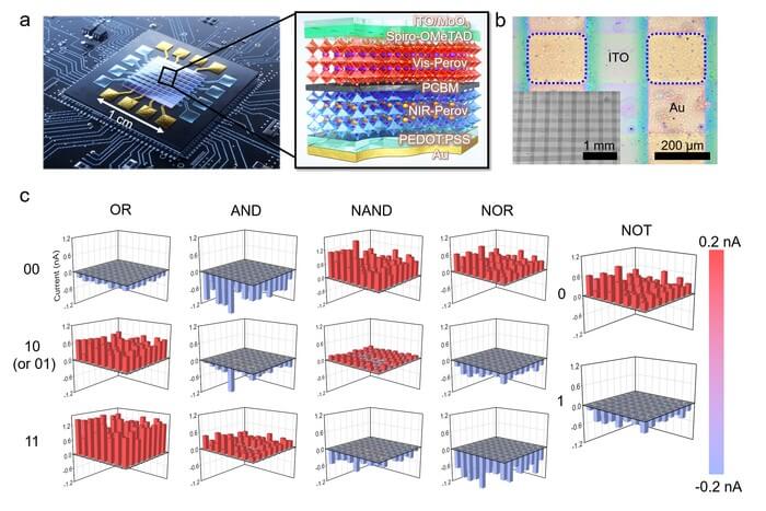
Three-dimensional bar charts for all the outputs (“OR”, “AND”, “NAND”, “NOR”, and “NOT”) obtained from the 64 pixels. The red and blue bars show clear bipolar spectral photoresponses of all the pixels with reference to the fiducial level of 0 nA (gray face).

The demand is explosively increasing for computers that can quickly calculate and process large amounts of information recently, as artificial intelligence, self-driving cars, drones, and metaverse technologies are drawing attention as core industries of the future. However, electronic semiconductor logic gates, which serve as the brains of computers today, have limited capacities in high-speed data calculation and processing and have disadvantages in that they consume a lot of energy and generate considerable heat.

Korea Institute of Science and Technology (KIST, President: Seok-Jin Yoon) and Gwangju Institute of Science and Technology (GIST, President: Ki-Seon Kim) announced that their research teams, led by Dr. Yusin Pak at the Sensor System Research Center (KIST) and Professor Gun Young Jung at the School of Materials Science and Engineering (GIST), have developed an ultra-high-speed, high-efficiency optoelectronic logic gates (OELGs) by using organic-inorganic perovskite photodiodes.

The optoelectronic logic gate has high-speed and high-efficiency characteristics; it uses light as an input signal which demonstrates low energy loss physically and can operate only with light energy without electrical power supply. The research teams implemented a stacked perovskite optoelectronic logic gate. Two layers of perovskite thin films are vertically stacked like a sandwich and proved that the desired binary logic operation is possible by inputting two lights of different wavelengths and intensities.

As the perovskite optoelectronic logic gate can freely change the photocurrent polarity using light, executing more than one logic gate operation result for the same input value is possible. Therefore, compared to the existing logic gate that can only perform one logical operation on one device, the newly developed one can implement all five different basic logic operations such as AND, OR, NAND, NOR, and NOT. It enables the development of optical processors with high spatial efficiency and integration, as one logic gate can function like five logic gates.

Dr. Pak (KIST) said, “Perovskite optoelectronic logic gates that execute multiple logic operations in response to optical input are expected to be used for ultra-small and low-power universal optical sensor platforms in the future.” Prof. Jung (GIST) expected that “The optoelectronic logic gate developed through this research is an outcome of optical computing R&D that realizes five basic logic operations into one device, and will greatly contribute to next-generation optical communication, optical network, and healthcare R&D”.

KIST was established in 1966 as the first government-funded research institute to establish a national development strategy based on science and technology and disseminate various industrial technologies to develop major industries. KIST is now raising Korean science and technology status through world-leading innovative research and development. For more information, please visit our website at https://eng.kist.re.kr/kist_eng_renew/

This study was supported by the KIST Institutional Program and the Mid-Career Researcher Program of the National Research Foundation of Korea (NRF) funded by the the Ministry of Science and ICT (Minister: Hye-Sook Lim). The research results were published in the Nature Communications, a renowned international journal (IF: 14.919, top 4.86% in JCR).


from ScienceBlog.com https://ift.tt/X5rhT4E

Does autism begin in the womb?

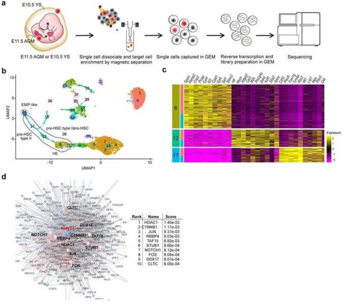
Fig. 1 Single-cell RNA-seq analysis of AGM. a: Experimental schematic. Cells are isolated from the fetal yolk sac and AGM and their transcripts are sequenced by next-generation sequencing. b: Cell groups from single-cell analysis of AGM progenitor cells are shown. c: Heatmap of top 10 enriched genes in the three cell groups (6, 12, 17) in b. d: Gene enrichment analysis based on protein-protein inter-network of RUNX1, a transcription factor essential for the endothelial-to-hematopoietic transition (EHT), in which vascular endothelial cells give rise to hematopoietic stem cells.

An international research group led by Professor Toru Takumi (Senior Visiting Scientist, RIKEN Center for Biosystems Dynamics Research) and Researcher Chia-wen Lin at Kobe University Graduate School of Medicine has shown that idiopathic autism*1 is caused by epigenetic*2 abnormalities in hematopoietic cells during fetal development, which results in immune dysregulation in the brain and gut. The results of the study revealed that in autism, there are immune abnormalities which can be seen in the brain and gut.

It is hoped that further classification of the pathophysiology of autism will lead to the creation of new treatment strategies for autism and other neurodevelopmental disorders*3.

The results of this research will be published in Molecular Psychiatry on Monday May 2, 2022 (1am BST).

Main Points

  • In BTBR mice*4, an animal model of autism, we identified HDAC1*5 as the etiology of immune abnormalities through single-cell RNA-seq*6 analysis of AGM*7 blood cell cells.
  • Single-cell RNA-seq analysis of yolk sac*8 hematopoietic cells also identified HDAC1 as the etiology of microglia*9 developmental abnormalities.
  • Regulation of HDAC activity during the fetal stage ameliorated inflammation in the brain and immune dysregulation in BTBR mice.
  • We found that changes in the intestinal environment, especially in the immune system, lead to abnormalities in the intestinal microbiota*10 of BTBR mice.

Research Background
Autism (autism spectrum disorder) is a developmental neurological disorder that remains largely unexplored despite the rapidly increasing number of patients. Immune abnormalities, now considered the cause of many diseases, also play an important role in the development of autism. Brain inflammation and disturbances of the peripheral immune system are frequently observed in autistic patients. Furthermore, immune abnormalities are accompanied by abnormalities in the intestinal microbiota, which is also thought to be involved in the pathogenesis of the disease via the brain-gut axis*11. However, the essential mechanisms behind these immune abnormalities have yet to be elucidated.

Given the critical developmental stages of immune insults and the extensive involvement of the immune system in the development of autism, the research team hypothesized that a common etiology underlies the widespread immune dysregulation and originates in different types of progenitor cells. The analysis focused on the hematopoietic cells from which immune cells are derived, as well as on the yolk sac (YS) and the aorta-gonad-mesonephros (AGM), which are involved in hematopoiesis during the fetal stage. These results seek a common ancestor of inflammation in the brain and abnormalities in the peripheral immune system. In this study, BTBR mice were used as an idiopathic model for autism.

Research Findings
Single-cell RNA sequencing (sc-RNA seq) of BTBR mice traced the origin of immune abnormalities back to the embryonic stages of the yolk sac (YS) and aorta-gonad-mesonephros (AGM) and identified where macrophages (microglia) and peripheral immune cells differentiate. Definitive hematopoiesis*12 in YS and AGM single-cell level analysis successfully identified pathological mechanisms at the molecular level within rare progenitor cells in the early stages of development. Namely, we found a common mechanism of transcriptional regulation through HDAC1, a histone deacetylase, underlying these pathologies (Figures 1 and 2).

We have also shown that manipulating epigenetic mechanisms during specific developmental stages can restore immune abnormalities in the brain and peripheral tissues. Namely, we identified histone deacetylase HDAC1 as a common mechanism. Administrating inhibitors of this histone (sodium butyrate or Romidepsin) during the fetal stage in BTBR mice suppressed elevated inflammatory cytokines*13 and microglial activation (Figure 3).

We further demonstrated that dysregulated immunity can determine gut dysbiosis of specific profiles in autistic model mice, which make the potential biomarkers of Treg and gut dysbiosis a means to categorize the immune-dysregulated ASD subtype.
From the above, it is clear that the abnormalities in the brain and peripheral organs (such as the intestines) seen in autism are caused by epigenetic abnormalities in the hematopoietic stem cell lineage, the ancestor of immune cells (Figure 4).

Perspectives
Our findings not only provide the missing piece to solve the long-time puzzle of systemic immune dysregulation in autism, but also hint the role of epigenetic disturbance as common etiology among different autism models of environmental risk factors. Furthermore, to develop precision medicine for ASD in the future, ASD subtyping according to the pathogenesis mechanism is a key first step to resolve the heterogeneity of ASD and to open up a new avenue for ASD treatment.

Glossary
*1 Idiopathic autism: Autism is considered to be a multifactorial disorder that can be caused by genetic and environmental factors. It is understood that genetic factors such as genetic and genomic abnormalities can cause autism, however there are still many cases of autism where the cause is unknown. Autism where the cause cannot be specified (including environmental factors) is called idiopathic autism.
*2 Epigenetics: The study of inheritance patterns that affect how genes work but do not involve alterations to the DNA sequence. Even though the information in the genome remains the same, biological mechanisms such as DNA methylation and chemical modification of histone proteins can alter genetic expression.
*3 Neurodevelopmental disorder:Previously called developmental disorders, this is a disorder that occurs in relation to a functional problem with the brain.
*4 BTBR mouse:A type of cogenic mouse. From analysis of the systemic behavior of this line of mice, it has been reported that BTBR mouse behavior is the closest to autistic behavior. Therefore, it is known as the idiopathic autism mouse model.
*5 HDAC1:Histone deacetylase 1 regulates gene expression by modifying histone proteins.
*6 Single-cell RNA-seq:A method of comprehensively investigating the qualitative and quantitative aspects of all mRNA present in individual cells using a next generation sequencer. By combining this with statistical analysis methods such as dimension reduction, it is possible to classify cells based on their genetic expression, and estimate the cell state. Furthermore, performing pseudo-temporal ordering analysis based on changes in the gene expression profile allows for the depiction of the fibers in the cellular state that accompanies development.
*7 AGM:The Aorta-gonad-mesonephros (AGM) region is a hematopoietic site within the fetus (i.e. where cellular components of the fetus’s blood are formed).
*8 Yolk sac:During pregnancy the sac, which is a membrane that surrounds the egg yolk, is also a hematopoietic site (primary hematopoiesis).
*9 Microglia: A type of glial cell in the central nervous system responsible for the central immune system. Microglia are also called the resident macrophages of the central nervous system. Unlike other glial cells (such as astrocytes and oligodendrocytes), microglia originate from yolk sac derived precursor cells.
*10 Intestinal microbiota: clusters of bacteria in the gut that are also called intestinal flora. Recent research advancements using a next-generation sequencer to analyze the metagenome of gut bacteria have shown links to various disorders including autism.
*11 Brain-gut axis: The relationship between the brain and the gut, also called the brain-gut connection. Two-way communication occurs between the brain and gut through mediums such as the autonomic nervous system and humoral factors (e.g. hormones and cytokines). Recently, this two-way communication system between the gut microbiome (microbiota) and the brain has received much attention.
*12 Definitive hematopoiesis: During the fetal period, hematopoiesis begins in the yolk sac with primary hematopoiesis and then secondary hematopoiesis occurs in the AGM region. Subsequent hematopoiesis during the fetal period occurs in the liver and lastly in the bone marrow. Hematopoiesis continues throughout a person’s life with bone marrow as the main site of this process.
*13 Inflammatory cytokine:A signaling molecule secreted by the immune cells, it causes inflammation.

Acknowledgements
This research received funding from sources including those listed below:

  • The following grants from the Japan Society for the Promotion of Science (JSPS): Grant-in-Aid for Scientific Research (S).
  • Japan Agency for Medical Research and Development’s Strategic Research Program for Brain Sciences
  • Research grant from the Takeda Science Foundation.

Journal Information
Title

“A common epigenetic mechanism across different cellular origins underlies systemic immune dysregulation in an idiopathic autism mouse model”
DOI:10.1038/s41380-022-01566-y

Authors
Chia-Wen Lin, Dian E Septyaningtrias, Hsu-Wen Chao, Mikiko Konda, Koji Atarashi, Kozue Takeshita, Kota Tamada, Jun Nomura, Yohei Sasagawa, Kaori Tanaka, Itoshi Nikaido, Kenya Honda, Thomas J McHugh, Toru Takumi

Journal
Molecular Psychiatry


from ScienceBlog.com https://ift.tt/cPDsi23

‘Eye-Catching’ Smartphone App Could Make It Easy To Screen for Neurological Disease at Home

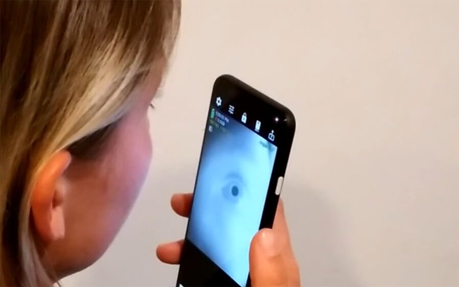
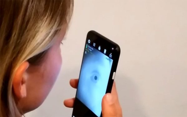
A smartphone user can image the eye using the RGB selfie camera and the front-facing near-infrared camera included for facial recognition. Measurements from this imaging could be used to assess the user’s cognitive condition. Credit: Digital Health Lab

A smartphone user can image the eye using the RGB selfie camera and the front-facing near-infrared camera included for facial recognition. Measurements from this imaging could be used to assess the user’s cognitive condition. Credit: Digital Health Lab

Researchers at the University of California San Diego have developed a smartphone app that could allow people to screen for Alzheimer’s disease, ADHD and other neurological diseases and disorders—by recording closeups of their eye.

The app uses a near-infrared camera, which is built into newer smartphones for facial recognition, along with a regular selfie camera to track how a person’s pupil changes in size. These pupil measurements could be used to assess a person’s cognitive condition.

The technology is described in a paper that will be presented at the ACM Computer Human Interaction Conference on Human Factors in Computing Systems (CHI 2022), which will take place from April 30 to May 5 in New Orleans as a hybrid-onsite event.

“While there is still a lot of work to be done, I am excited about the potential for using this technology to bring neurological screening out of clinical lab settings and into homes,” said Colin Barry, an electrical and computer engineering Ph.D. student at UC San Diego and the first author of the paper, which received an Honorable Mention for Best Paper award. “We hope that this opens the door to novel explorations of using smartphones to detect and monitor potential health problems earlier on.”

Pupil size can provide information about a person’s neurological functions, recent research has shown. For example, pupil size increases when a person performs a difficult cognitive task or hears an unexpected sound.

Measuring the changes in pupil diameter is done by performing what’s called a pupil response test. The test could offer a simple and easy way to diagnose and monitor various neurological diseases and disorders. However, it currently requires specialized and costly equipment, making it impractical to perform outside the lab or clinic.

Engineers in the Digital Health Lab, led by UC San Diego electrical and computer engineering professor Edward Wang, collaborated with researchers at the UC San Diego Center for Mental Health Technology (MHTech Center) to develop a more affordable and accessible solution.

“A scalable smartphone assessment tool that can be used for large-scale community screenings could facilitate the development of pupil response tests as minimally-invasive and inexpensive tests to aid in the detection and understanding of diseases like Alzheimer’s disease.  This could have a huge public health impact,” said Eric Granholm, a psychiatry professor at UC San Diego School of Medicine and director of the MHTech Center.

The app developed by the UC San Diego team uses a smartphone’s near-infrared camera to detect a person’s pupil. In the near-infrared spectrum, the pupil can be easily differentiated from the iris, even in eyes with darker iris colors. This enables the app to calculate pupil size with sub-millimeter accuracy across various eye colors. The app also uses a color picture taken by the smartphone’s selfie camera to capture the stereoscopic distance between the smartphone and the user. The app then uses this distance to convert the pupil size from the near-infrared image into millimeter units.

The app’s measurements were comparable to those taken by a device called a pupillometer, which is the gold standard for measuring pupil size.

The researchers also included various features in their app to make it more user friendly for older adults.

“For us, one of the most important factors in technology development is to ensure that these solutions are ultimately usable for anyone. This includes individuals like older adults who might not be accustomed to using smartphones,” said Barry.

The researchers worked with older adult participants to design a simple app interface that allows users to self administer pupil response tests. This interface included voice commands, image-based instructions, and a cheap, plastic scope to direct the user to place their eye within the view of the smartphone camera.

“By testing directly with older adults, we learned about ways to improve our system’s overall usability and even helped us innovate older adult specific solutions that make it easier for those with different physical limits to still use our system successfully,” said Wang, who is also a faculty member in the UC San Diego Design Lab. “When developing technologies, we must look beyond function as the only metric of success, but understand how our solutions will be utilized by end-users who are very diverse.”

The Digital Health Lab is continuing this work in a project to enable similar pupillometry function on any smartphone rather than just the newer smartphones. Future studies will also involve working with older adults to evaluate home use of the technology. The team will work with older individuals with mild cognitive impairment to test the app as a risk screening tool for early stage Alzheimer’s disease.

This work was funded by the National Institute of Aging.

To learn more about this project and other works by the UCSD Digital Health Lab, see their webpage at https://digihealth.eng.ucsd.edu/.

Paper: “At-Home Pupillometry using Smartphone Facial Identification Cameras.


from ScienceBlog.com https://ift.tt/WCD0Jr3

‘Resetting’ the injured brain offers clues for concussion treatment

New research in mice raises the prospects for development of post-concussion therapies that could ward off cognitive decline and depression, two common conditions among people who have experienced a moderate traumatic brain injury.

The study in mice clarified the role of specific immune cells in the brain that contribute to chronic inflammation. Using a technique called forced cell turnover, researchers eliminated these cells in the injured brains of mice for a week and then let them repopulate for two weeks.

“It’s almost like hitting the reset button,” said senior study author Jonathan Godbout, professor of neuroscience in The Ohio State University College of Medicine.

Compared to brain-injured mice recovering naturally, mice that were given the intervention showed less inflammation in the brain and fewer signs of thinking problems 30 days after the injury.

Though temporarily clearing away these cells, called microglia, in humans isn’t feasible, the findings shed light on pathways to target that could lower the brain’s overall inflammatory profile after a concussion, potentially reducing the risk for behavioral and cognitive problems long after the injury.

“In a moderate brain injury, if the CT scan doesn’t show damage, patients go home with a concussion protocol. Sometimes people come back weeks, months later with neuropsychiatric issues. It’s a huge problem affecting millions of people,” said Godbout, faculty director of Ohio State’s Chronic Brain Injury Program and assistant director of basic science in the Institute for Behavioral Medicine Research.

“How do you treat that? At least in mice, by turning over the microglia in the brain we had a very positive effect on their behavior, cognitive status and level of inflammation in the brain. Now we can focus on cellular pathways that generate chronic inflammation as a target.”

The research is published online in the Journal of Neuroscience.

About 85% of traumatic brain injuries are similar to the type of concussion examined in this study, involving dispersed impact to the head that causes brain tissue to bump against the skull. Previous research suggests that at least 75% of people who experience a moderate brain injury have long-term mental health and cognitive complications.

Godbout’s lab previously linked depressive symptoms in mice to microglia’s sustained “high alert” status after a head injury, which causes the cells to overreact to later challenges to the immune system and become excessively inflammatory. In a more recent study in mice, his team showed that forced turnover of microglia before a head injury could reduce later neuropsychiatric complications.

“That was a proof of principle to show that a lot of the inflammation, especially in the long term, is mediated by microglia,” he said. “But there is an acute phase of inflammation – you want to initiate that repair process. There’s a positive to that early inflammatory response in the brain or spinal cord. If it lasts a long time and doesn’t fully resolve, that’s when it’s dangerous.

In this new study, researchers waited for seven days after the brain injury to force the turnover of microglia, giving the cells time to carry out their work promoting initial healing. An experimental drug that inhibits a protein that microglia in mice need for survival was added to their food for a week, resulting in depletion of over 95% of microglia in their brains.

After allowing 16 days for the microglia to repopulate, researchers compared the intervention mice to injured mice that recovered without the cell turnover treatment. The intervention mice performed better than control mice on tasks testing their memory and depressive symptoms.

Further analyses of injured brain tissue suggested the cell turnover reversed some injury-related damage to neurons, lowered overall inflammation and improved the brain’s ability to adapt to change. Researchers also injected mice with a molecule that triggers an immune response to mimic an infection, and found that sickness behavior was lower in the intervention mice.

Godbout said these combined findings suggest that the repopulating microglia returned in a less “primed” state of readiness, lowering chances for a lifetime of exaggerated inflammatory responses in the brain to any challenge to the immune system – that brain inflammation being the likely culprit behind the neuropsychiatric complications that follow a head injury.

“If microglia in the human brain don’t return to normal and chronic inflammation persists after a head injury, it’s not just a secondary brain injury that causes problems. Even getting a viral infection after concussion recovery can progress into a cognitive or behavioral issue or amplify some other part of behavior, like depression,” Godbout said. “There is a real connection between a head injury and mental health, and the risk doesn’t go away.

“Now we’re looking more closely at the pathways that cause changes in microglia, and targeting something specific in that pathway. That is a way forward.”

This work was supported by the National Institute of Neurological Disorders and Stroke, the National Institute on Aging, the National Institute of Dental and Craniofacial Research, an Ohio State University Presidential Fellowship and the Thailand Research Fund-Royal Golden Jubilee Program.

Co-authors include Chelsea Bray, Kristina Witcher, Dunni Adekunle-Adegbite, Michelle Ouvina, Mollie Witzel, Emma Hans, Zoe Tapp, Jonathan Packer, Ethan Goodman, Fangli Zhao, Shane O’Neil, John Sheridan, Olga Kokiko-Cochran and Candice Askwith, all of Ohio State, and Titikorn Chunchai and Siriporn Chattipakorn of Chiang Mai University in Thailand.


from ScienceBlog.com https://ift.tt/4EkwWp2

Organic pesticides to provide natural protection for endangered crops

Some vitally important European crops like vines and olives are being devastated by disease. Scientists are searching for biological replacements for chemical pesticides to improve crop and human health.

The threat to agriculture from invasive species is huge. The United Nations (UN) estimates that plant disease costs the world’s economy over €200 billion per year, with 20-40% of crop production lost to pests.

‘The economic loss from invasive species is immense, and if we took no action, there would be a huge amount of food insecurity, not only across the EU but across the globe,’ said Dr. Hikmate Abriouel, professor of microbiology at Universidad de Jaén in Spain’s Andalucía.

With the stakes so high, it’s easy to understand why the agricultural sector is one of the largest users of chemicals worldwide.

The question of food security is uppermost these days. But, as Dr. Abriouel goes on to explain, our growing reluctance to use chemicals in agriculture adds a layer of complication to farming.

‘There was a time when it was normal to rely on powerful pesticides to treat agricultural land,’ she said. ‘But now we know that a chemical designed to kill a living organism is likely to have negative impacts on other biological systems too.’

Spraying crops with synthetic compounds has adverse impacts on people, farm animals, wildlife, pollinators like bees and other living things that play an essential role in the ecosystem. The chemical runoff also damages the land and water.

Pollution risk

Pesticide pollution causes risk to farmland from the chemical residues that leach into water supplies.

Some synthetic pesticides have been linked to human diseases like cancer, diseases of the immune system and respiratory illnesses.

Farmers who work with pesticides are particularly vulnerable to side-effects, with an estimated 44% of farm workers worldwide experiencing at least one incident of acute pesticide poisoning every year.

The EU’s Farm to Fork (F2F) strategy for sustainable food production targets significant reductions in the use of chemical pesticides, fertilisers and antimicrobials and supports an increase in organic farming. Sustainability goals mean biopesticides or biological alternatives to pesticides are required.

‘There is a lot of evidence that replacing chemicals with biopesticides works with nature rather than against it,’ said Dr. Abriouel. Biological solutions benefit soil health and biodiversity too.

Dying vines

In France alone, around 12% of vineyards were unproductive between 2012 and 2017 due to Grape Trunk Disease (GTD) which has been spreading across Europe over the past two decades. A chemical pesticide used to treat vines was banned because it is harmful to human and environmental health.

The disease results in 50% less productive plants, a decrease in the quality of the wine and the premature death of healthy vines. Worldwide, estimates for the replacement cost of grapevines exceed €1.4 billion per year.

As a response to this blight, the EU is funding the multinational BIOBESTicide project which aims to find a biological solution to GTD.

‘Our aim is to produce a really effective, totally natural preventive solution to this very serious and very expensive problem,’ said Dr. Assia Dreux-Zigha who works for the French biotechnology company Greencell and is coordinating the BIOBESTicide research.

The team’s research is focused on a specific strain of Pythium oligandrum – a ‘friendly’ fungus that is naturally present in the rhizosphere of many crop plants, including vines. The rhizosphere is the microorganism-rich region of soil directly around a plant’s roots.

P. oligandrum works both by destroying parasites directly and by inducing plant resistance against further attack. After isolating P. oligandrum in the lab, Greencell and its partners found that under certain conditions, the biopesticide colonised the roots of vines and stimulated the plant’s natural defences against GTD.

In the near future, following trials and safety approval, the BIOBESTicide researchers aim to scale up and field-test their biopesticide in vineyards across different geographical areas.

‘This is a very challenging project but, when we finish in late-2023, we hope to have a solution that will make it possible for vine plants to survive for their entire natural lifecycles,’ said Dr. Dreux-Zigha.

Undoubtedly, winemakers will raise a glass to this prospect.

Olive preserver

A second iconic European crop urgently in need of a biopesticide solution is the olive. First detected in European olives in 2013, Olive Quick Decline Syndrome (OQDS) is the disease caused by the bacterium Xylella fastidiosa.

In Puglia, southern Italy, where Xylella first surfaced on the continent, olive production shrank by 65-80% in the years up to 2020 with the loss of an estimated 100 000 jobs and the destruction of 400-year-old heritage olive trees.

Xylella has surfaced in France, Spain and Portugal, spread by an insect called the spittlebug. Affected plants are infected from the roots upwards, causing the leaves to turn brown and eventually killing the plant. It is considered one of the most dangerous plant pathogenic bacteria in the world.

‘The problem with this pathogen is getting worse,’ said Dr. Abriouel, who supervises the EU-backed SMART-AGRI-SPORE project, which aims to develop a biopesticide based on bacterial spores.

‘Preventing further spread of this pest is a priority in the EU,’ she said. A 2020 study estimated that as a worst-case scenario, Italy alone stands to lose between €1.9 billion and €5.2 billion over a 50-year period as a result of OQDS.

A number of projects are developing biopesticides to attack Xylella. Principal researcher Dr. Julia Manetsberger under the supervision of Dr. Abriouel is focused on modifying a strain of another bacteria to render it deadly to Xylella.

The researchers are hopeful that by 2024, a viable biopesticide will emerge from this research.

‘We can’t use something against Xylella that changes the biodiversity or destroys or increases the resistance of microorganisms present in other plants and soil,’ said Dr. Abriouel. ‘In other words, we can’t solve one problem and create another.’

‘We are working hard to reach this objective,’ said Dr. Manetsberger, ‘These plants are so important for our economy and we need to defend them.’

The research in this article was funded by the EU. If you liked this article, please consider sharing it on social media.


from ScienceBlog.com https://ift.tt/pe9P3UJ

Search reveals eight new sources of black hole echoes

Scattered across our Milky Way galaxy are tens of millions of black holes — immensely strong gravitational wells of spacetime, from which infalling matter, and even light, can never escape. Black holes are dark by definition, except on the rare occasions when they feed. As a black hole pulls in gas and dust from an orbiting star, it can give off spectacular bursts of X-ray light that bounce and echo off the inspiraling gas, briefly illuminating a black hole’s extreme surroundings.

Now MIT astronomers are looking for flashes and echoes from nearby black hole X-ray binaries — systems with a star orbiting, and occasionally being eaten away by, a black hole. They are analyzing the echoes from such systems to reconstruct a black hole’s immediate, extreme vicinity.

In a study appearing today in the Astrophysical Journal, the researchers report using a new automated search tool, which they’ve coined the “Reverberation Machine,” to comb through satellite data for signs of black hole echoes. In their search, they have discovered eight new echoing black hole binaries in our galaxy. Previously, only two such systems in the Milky Way were known to emit X-ray echoes.

In comparing the echoes across systems, the team has pieced together a general picture of how a black hole evolves during an outburst. Across all systems, they observed that a black hole first undergoes a “hard” state, whipping up a corona of high-energy photons along with a jet of relativistic particles that is launched away at close to the speed of light. The researchers discovered that at a certain point, the black hole gives off one final, high-energy flash, before transitioning to a “soft,” low-energy state.

This final flash may be a sign that a black hole’s corona, the region of high-energy plasma just outside a black hole’s boundary, briefly expands, ejecting a final burst of high-energy particles before disappearing entirely. These findings could help to explain how larger, supermassive black holes at the center of a galaxy can eject particles across vastly cosmic scales to shape a galaxy’s formation.

“The role of  black holes in galaxy evolution is an outstanding question in modern astrophysics,” says Erin Kara, assistant professor of physics at MIT. “Interestingly, these black hole binaries appear to be ‘mini’ supermassive black holes, and so by understanding the outbursts in these small, nearby systems, we can understand how similar outbursts in supermassive black holes affect the galaxies in which they reside.”

The study’s first author is MIT graduate student Jingyi Wang; other co-authors  include Matteo Lucchini and Ron Remillard at MIT, along with collaborators from Caltech and other institutions.

X-ray delays

Kara and her colleagues are using X-ray echoes to map a black hole’s vicinity, much the way that bats use sound echoes to navigate their surroundings. When a bat emits a call, the sound can bounce off an obstacle and return to the bat as an echo. The time it takes for the echo to return is relative to the distance between the bat and the obstacle, giving the animal a mental map of its surroundings.

In similar fashion, the MIT team is looking to map the immediate vicinity of a black hole using X-ray echoes. The echoes represent time delays between two types of X-ray light: light emitted directly from the corona, and light from the corona that bounces off the accretion disk of inspiraling gas and dust.

The time when a telescope receives light from the corona, compared to when it receives the X-ray echoes, gives an estimate of the distance between the corona and the accretion disk. Watching how these time delays change can reveal how a black hole’s corona and disk evolve as the black hole consumes stellar material.

Echo evolution

In their new study, the team developed search algorithm to comb through data taken by NASA’s Neutron star Interior Composition Explorer, or NICER, a high-time-resolution X-ray telescope aboard the International Space Station. The algorithm picked out 26 black hole X-ray binary systems that were previously known to emit X-ray outbursts. Of these 26, the team found that 10 systems were close and bright enough that they could discern X-ray echoes amid the outbursts. Eight of the 10 were previously not known to emit echoes.

“We see new signatures of reverberation in eight sources,” Wang says. “The black holes range in mass from five to 15 times the mass of the sun, and they’re all in binary systems with normal, low-mass, sun-like stars.”

As a side project, Kara is working with MIT education and music scholars, Kyle Keane and Ian Condry, to convert the emission from a typical X-ray echo into audible sound waves. Take a listen to the sound of a black hole echo here:

The researchers then ran the algorithm on the 10 black hole binaries and divided the data into groups with similar “spectral timing features,” that is, similar delays between high-energy X-rays and reprocessed echoes. This helped to quickly track the change in X-ray echoes at every stage during a black hole’s outburst. 

The team identified a common evolution across all systems. In the initial “hard” state, in which a corona and jet of high-energy particles dominates the black hole’s energy, they detected time lags that were short and fast, on the order of milliseconds. This hard state lasts for several weeks. Then, a transition occurs over several days, in which the corona and jet sputter and die out, and a soft state takes over, dominated by lower-energy X-rays from the black hole’s accretion disk.   

During this hard-to-soft transition state, the team discovered that time lags grew momentarily longer in all 10 systems, implying the distance between the corona and disk also grew larger. One explanation is that the corona may briefly expand outward and upward, in a last high-energy burst before the black hole finishes the bulk of its stellar meal and goes quiet.

“We’re at the beginnings of being able to use these light echoes to reconstruct the environments closest to the black hole,” Kara says. “Now we’ve shown these echoes are commonly observed, and we’re able to probe connections between a black hole’s disk, jet, and corona in a new way.”

This research was supported, in part, by NASA.


from ScienceBlog.com https://ift.tt/crhNRim

Featured Post

Show your sources: building verifiable, citable AI agents with MCP

Model Context Protocol (MCP) is an open standard which connects LLMs with external systems. We discuss how new Dimensions and Altmetric MCP...

Popular